肝血窦
外观
| 肝血窦 | |
|---|---|
 | |
 Basic liver structure | |
| 标识字符 | |
| 拉丁文 | vas sinusoideum |
| TH | H3.04.05.0.00014 |
| FMA | FMA:17543 |
| 《解剖學術語》 [在维基数据上编辑] | |
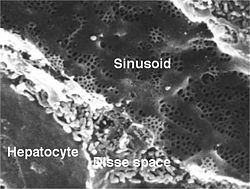
肝血窦(英语:Liver sinusoid或Hepatic sinusoids)是一种被称为窦状毛细血管、不连续毛细血管或血窦的毛细血管。它类似于有孔毛细血管,具有不连续的内皮细胞,可作为来自肝总动脉的富氧血液和来自肝门静脉的富营养血液的混合位置。[1]
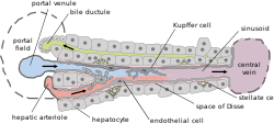
肝血窦具有比其他类型的毛细血管更大的口径,并具有称为肝窦内皮细胞(LSEC)和库普弗细胞的特化内皮细胞衬里。[2]这些细胞是多孔的并且具有清除功能。[3]LSEC约占肝脏中非实质细胞的一半,扁平且有孔,[4]可以使窦腔和窦周间隙之间更容易交流。LSEC和库普弗细胞在过滤、胞吞作用和调节血窦中的血流方面发挥作用。[5]
库普弗细胞可以吞食外来物质,例如细菌。窦周间隙将肝细胞与血窦分开。肝脏星状细胞存在于窦周间隙中,并参与对肝脏的纤维化反应。
当LSEC丢失使窦道成为一个普通的毛细血管时,就会发生剥落现象。这一过程先于纤维化发生。[6]
内皮细胞
[编辑]肝血窦内皮细胞可用于各种研究目的。这些细胞的效用特别令人感兴趣。只是要克服它的一个问题,那就是细胞分化的逆转。这种逆转使这些细胞在体外表型上高度特化。[7]
图像
[编辑]-
人类肝血窦
-
猪肝脏的单个小叶
参考文献
[编辑]- ^ SIU SOM Histology GI. [2023-01-28]. (原始内容存档于2017-07-17).
- ^ Brunt, EM; et al. Pathology of the liver sinusoids.. Histopathology. June 2014, 64 (7): 907–20. PMID 24393125. S2CID 12709169. doi:10.1111/his.12364.
- ^ DeLeve, LD. Hepatic microvasculature in liver injury.. Seminars in Liver Disease. November 2007, 27 (4): 390–400. PMID 17979075. doi:10.1055/s-2007-991515.
- ^ Xing, Y; Zhao, T; Gao, X; Wu, Y. Liver X receptor α is essential for the capillarization of liver sinusoidal endothelial cells in liver injury.. Scientific Reports. 18 February 2016, 6: 21309. Bibcode:2016NatSR...621309X. PMC 4758044
 . PMID 26887957. doi:10.1038/srep21309.
- ^ Arii, S; Imamura, M. Physiological role of sinusoidal endothelial cells and Kupffer cells and their implication in the pathogenesis of liver injury.. Journal of Hepato-biliary-pancreatic Surgery. 2000, 7 (1): 40–8. PMID 10982590. doi:10.1007/s005340050152.
- ^ Xie, G; Wang, X; Wang, L; Wang, L; Atkinson, RD; Kanel, GC; Gaarde, WA; Deleve, LD. Role of differentiation of liver sinusoidal endothelial cells in progression and regression of hepatic fibrosis in rats.. Gastroenterology. April 2012, 142 (4): 918–927.e6. PMC 3618963
 . PMID 22178212. doi:10.1053/j.gastro.2011.12.017.
- ^ Sellaro TL, Ravindra AK, Stolz DB, Badylak SF. Maintenance of hepatic sinusoidal endothelial cell phenotype in vitro using organ-specific extracellular matrix scaffolds. Tissue Eng. September 2007, 13 (9): 2301–2310 [28 April 2013]. PMID 17561801. doi:10.1089/ten.2006.0437.
外部链接
[编辑]- UIUC組織學主題 589
- Histology image: 15504loa – 波士顿大学的组织学学习系统 - "Liver, Gall Bladder, and Pancreas: liver, classic lobule"
- Histology image: 22103loa – 波士顿大学的组织学学习系统 - "Ultrastructure of the Cell: hepatocytes and sinusoids, sinusoid and space of Disse"
- Histology at anhb.uwa.edu.au (页面存档备份,存于互联网档案馆)


